News reporting by Aarya Krishnan.
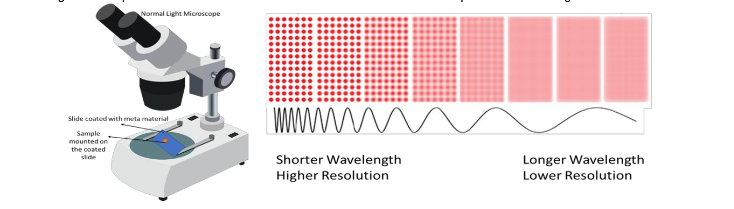
San Diego, June 1: A light microscope can be converted into a high-resolution microscope thanks to a new metamaterial developed by researchers at the University of California, San Diego. Metamaterials are artificially engineered materials on the microscale or nanoscale that can achieve extraordinary properties. This particular metamaterial is made up of nanometer-thin silver and silica glass layers which alternate with each other. Because of the specific arrangement of the layers, the wavelength of light is reduced as it passes through this substance. This is a very helpful characteristic in microscopy since shorter wavelengths imply higher resolution.
The resolution of a microscope refers to its capacity to distinguish two objects distinctly as independent objects under a microscope. The wavelength of the light used to see the sample influences the resolution. A microscope’s resolution increases as the wavelength decreases. Although light microscopes are excellent for studying live cells, their resolution is just 200 nanometers(nm). This makes light microscopes ineffective for imaging subcellular structures (smaller components of a cell). Despite the fact that powerful microscopes, such as electron microscopes, provide higher resolution for seeing subcellular characteristics, they cannot be used to image living cells since the samples must be placed inside a vacuum chamber.

Imaging subcellular structures in living cells can be a useful tool for learning more about the biological mechanisms at work. When a sample is placed on a microscope slide coated with metamaterial (Picture 1), the wavelength of the light that travels through the metamaterial shortens before it illuminates the sample, allowing the microscope to photograph the sample with higher resolution (Picture 2). The resolution was raised to 40nm, enabling for the imaging of finer features like actin filaments that would be difficult to see with simply the microscope. Because the microscope does not require any specific modifications, it is a particularly appealing solution to the problem of low resolution. Their original work was published in Nature Communications.